Ophtalmic Ultrasound Use in Veterinary medicine
In most circumstances the retina can be visualized during a complete ophthalmic examination. In patients in which this is not possible, ophthalmic ultrasound can provide a detailed picture of the retina using ophtalmic ultrasound. Because the eye is a superficial fluid filled structure, ultrasound is an easy to use modality for visualization of ocular pathology and anatomy. Topical anesthetic is administered to the surface of the eye, and ultrasound gel is then applied to the eyelids. The ultrasound transducer is then placed on the outside of the eyelids and a detailed picture of the interior of the eye is obtained.
The most imporant goal of ultrasound is to detect a retinal detachment. This step is necessary before cataract surgery. Patients with retinal detachments are not elligible for this procedure. There are conditions besides cataracts in which the retina cannot be visualized, and in these cases, Ophtalmic ultrasound can be a valuable tool in determining whether or not a retinal detachment is present. This is crucial in determining treatment and prognosis.
The principles of ocular ultrasound are similiar in concept to other applications of this technology. Sound waves are produced at a frequency higher than 20,000 Hz (20 kHz) and reflected back to the transducer by tissue in its path. When the sound wave returns, a piezo-electric crystal in the transducer vibrates, producing an electrical signal that are converted into an image or other data.
Higher frequency has more shallow penetration into tissue but have better resolution. In contrast, lower frequency waves penetrate more deeply but have worse resolution. Ultrasound waves, like other waves, have predictive behaviors based on properties of the medium they travel through. For instance, sound waves have higher velocity when traveling through solids than through liquids. When sound waves travel between tissue interfaces with different acoustic impedance, or densities, they can either scatter, reflect, or refract. Some sound is absorbed by tissue as well. Sound waves that return to the transducer are called echoes, and ultrasound imaging zones can be hyperechoic, hypoechoic, or anechoic. Shadowing can occur distal to a very dense lesion, resulting in an anechoic region.
There are two main types of ultrasound used in ophthalmologic practice currently, A-Scan and B-scan. In A-scan, or time-amplitude scan, sound waves are generated at lower ultrasound wavelength and converted into spikes that correspond with tissue interface zones. In B-scan, or brightness amplitude scan, sound waves are generated at higher ultrasound wavelength. The data collected by the transducer produces a corresponding image.
A-scans in veterinary medicine are typically used for biometry, or measurement of ocular structures. Exams can be performed in awake, sedated or anesthetized animals. The probe can be placed directly on the cornea, or used with a scleral shell and waterbath (immersion technique). The probe should always be placed axially. A good scan is one in which the height of the spikes from baseline are equal. Each spike should start at a perpendicular, not sloping angle from baseline. A-scan has not been used to any great extent in veterinary medicine. One reason for this is that diagnostic A-scans are used to diagnose choroidal tumors, which are much more common in humans than in the typical veterinary patient. The anterior uveal tumors more common in veterinary medicine are harder to image with A-scan. This makes the A-scan only useful for biometry and measurement of occular structures in veterinary feild.
B-scan type of ophtalmic ultraasound typically used for evaluation of intraocular structures that cannot be seen through opaque media, such as corneal opacities, hemorrhage or hypopyon in the anterior chamber, cataracts, or vitreal opacities. In veterinary medicine, the most common probe position is axial. The scan is performed with the eye in primary gaze and the probe face centered on the cornea. The image is intersected by the optic nerve as the sound beam is directed through the center of the lens, and the beam is swept along the two opposing meridians. This image is usually easiest to understand because the lens and optic nerve are in the center of the lesion, but there is decreased resolution of the posterior segment due to sound attenuation and refraction from the lens. In veterinary medicine, however, this is the easiest probe position to use in conscious animals.
The recent development of a 20-MHz, high-frequency ultrasound probe has allowed tissue to be visualized at resolutions of 20 to 80 microm, which is similar to a low-power histologic view. This high degree of resolution, however, limits tissue penetration to 5 to 10 mm, which is ideal for examination of the anterior segment of the eye. The detail provided by high-resolution ultrasound readily permits the clinician to distinguish between various anterior segment entities that may appear similar but are treated quite differently, such as anterior uveal tumors, iridociliary cysts, and iris bombé. High-frequency ultrasound is also a valuable aid in creating a surgical plan for treatment of ocular disorders in which the cornea is opaque, such as feline corneal sequestrum and tumor invasion into the cornea. Other applications of this technology include elucidation of the pathogenesis of glaucoma in veterinary patients and evaluation of regions of the lens that are difficult to examine directly.
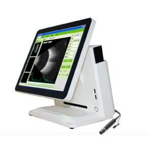
When it comes to ophtalmic ultrasound procedures where there is no cataract to hinder the rsolution, the ophtalmic probe ultrasound scanner SIFULTRAS-8.25 is suitable for measuring the anterior chamber depth, lens thickness, vitreous body length, axial length and calculating the IOL power for an implanted lens. This Ocular Ultrasound Probe thanks to it’s high frequency resulution (20MHz) can provide accurate measurement of central and peripheral corneal thickness and, is widely used in the preoperative examination and postoperative effect evaluation of refractive surgery.
In other circumstance there can an obstruction hindering the resolution and accuracy of the ophtalmic ultrasound reading such as cataract, or genetic defects. In this case the Ophthalmic Ultrasound Scanner SIFULTRAS-8.1 A/B Scan is best recommended for Ocular B-mode ultrasonography as one of the noninvasive, rapid diagnostic imaging techniques indicated in cataract patients to evaluate the posterior segment of the eye. This Ophthalmic A/B Scanner with normal, vitreous body enhancement, retina observation mode is mainly used for diagnosis of intraocular diseases, display the location, shape range of the focus of infection and the relationship with the surrounding tissue. It can diagnose vitreous opacity, retinal detachment, eye base tumors etc.
These procedures are performed by a certified veterinary ophtalmologist*
Reference: Ophthalmologic Ultrasound
Ocular ultrasonographic evaluation of cataractous and pseudophakic eyes in dogs