

Advantages of Pediatric Ultrasound Imaging
Ultrasound is of high clinical value and high accuracy in the diagnosis of pediatric abdominal pain. The etiology of pediatric abdominal pain has a certain specificity. Ultrasound can not only diagnose the cause of abdominal pain in pediatric patients, but also help clinicians to formulate the correct treatment plan. Ultrasound examination has the advantages of fast, simple, safe and feasible, no radiation radiation, and accurate diagnosis. It is the first choice for the diagnosis of pediatric abdominal pain.
Ultrasound is the most important imaging technique in pediatrics, more so than in any other branch of medicine. This is attributs to a number of factors. First of all, X-ray exposure is a more significant risk factor for pediatric patients than for adults by virtue of the cumulative nature of X-ray dosages, thus making paediatric patients more likely to receive a higher dose of radiation. Moreover, some types of immature tissue are more susceptible to radiation. Thus it is essential to avoid the use of X-rays in children and young adults.
Second of all, various types of heart catheterisation can be done using ultrasound instead of radiography. In addition, complex imaging of skull pathology in paediatric patients can be realised using ultrasound rather than radiography.
Which ultrasound scanner is best for pediatrics?
The SIFULTRAS-5.45. Note that the higher frequency peak of up to 70 MHz provides a tremendous increase in resolution compared with that of conventional ultrasound systems, permitting resolution down to 30 μm. Such resolution surpasses that of CT or MRI. Note that an increased frequency results in an increase in ultrasound attenuation in tissues, which means that the use of the system is limited to the first 3 cm of the body. Such resolution can be used to depict single hair follicles growing out of the skin.
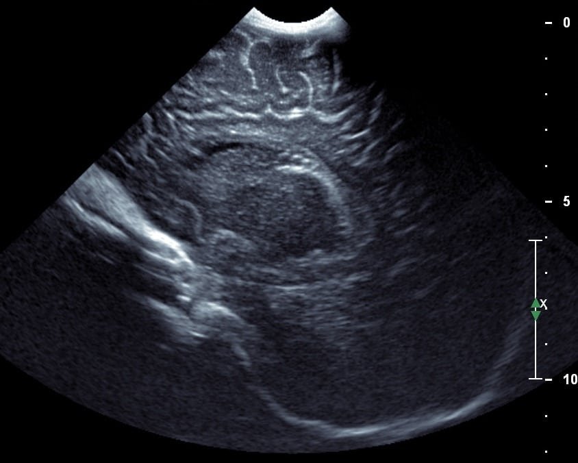
The High Frequency ultrasound scanner SIFULTRAS-5.45 heads (ideal for neonatology by virtue of their relatively shallow penetration depth) often produce image quality that is unmatched by any other method. These type of ultrasound scanner offer High resolution. In some cases, only ultrasound can detect congenital heart defects, brain morphology defects, cranial bleeding, and haemodynamic complications. Although MRI provides outstanding image quality in infants, the examinations are lengthy and an infant must be either anaesthetised or heavily sedated.
Ultrasound is widely available and can be used for screening purposes. For example, the current practice of doing a hip screening for all newborns has reduced to a minimum the number of hip defects requiring surgery.
A variety of pediatric venous diseases leading to the formation of debris or thrombus in the superficial and deep veins can be evaluated with high-resolution ultrasound. Other applications include improved characterization of nerve fascicles, large- and small-vessel intima-media thickness, internal contents of peripheral veins, and salivary glands. Thyroid nodules, superficial vascular anomalies, and ocular pathology, especially in small children, would also be better evaluated with high-resolution imaging.
It is important to note that for evaluation of superficial soft-tissue abnormalities in small children, ultrasound provides higher spatial resolution than MRI. The spatial resolution of a high-resolution ultrasound scanner is in micrometers, whereas that of MRI is in millimeters. With the availability of FDA-approved ultrasound contrast, ultrasound can now provide dynamic enhancement information similar to multiphase contrast-enhanced MRI.
Moreover, sedation required for MRI of most small children is not without risks. Although the diagnostic benefits of MRI should be acknowledged, the awareness that, in some circumstances, advanced ultrasound techniques can provide the necessary diagnostic information is worth noting. In referral of pediatric patients to radiologic exams, such knowledge would be beneficial.
Reference: The advantages of ultrasound for paediatric diagnosis.

[launchpad_feedback]
Disclaimer: Although the information we provide is used by different doctors and medical staff to perform their procedures and clinical applications, the information contained in this article is for consideration only. SIFSOF is not responsible neither for the misuse of the device nor for the wrong or random generalizability of the device in all clinical applications or procedures mentioned in our articles. Users must have the proper training and skills to perform the procedure with each vein finder device.
The products mentioned in this article are only for sale to medical staff (doctors, nurses, certified practitioners, etc.) or to private users assisted by or under the supervision of a medical professional.
