

Ultrasound-guided Vascular Cannulation
Traditional anatomical landmark technique for vascular cannulation, have been a source for various failures and complications.
On the other hand, ultrasound-guided vascular cannulation has been shown to improve first pass success, reduce the number of attempts, improve patient’s satisfaction and reduce overall procedure-related complications.
Which ultrasound scanner is best for vascular cannulation?
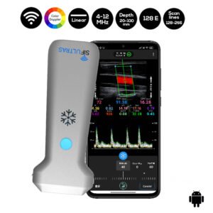
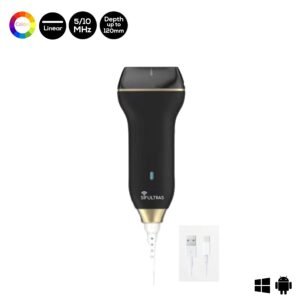
Since vascular structures are superficial, a linear array transducer (7–10MHz) SIFULTRAS-5.34 is commonly used to recognize the vessels, with the selection of a preset corresponding to veins or arteries, as the case may be.
Ultrasound-guided vascular cannulation is becoming the modality of choice to obtain tool to obtain a secure vascular access in critical care patients.
Depth, gain and focal zones are the most important machine parameters that practitioners always need to optimize to carry out the best possible evaluation on the vessels. Preferably, tissue harmonic imaging must be switched off, especially for further needle recognition.
Nonetheless, to summarize and simplify the fundamental sonographic aspects at the moment of deciding and executing an ultrasound-guided vascular access, and it also intends to highlight some aspects related to the optimal training for this practice. It goes as follows;
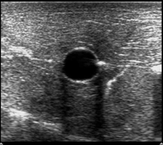
After recognizing the vascular structures, the next step is selecting an adequate vessel to be successfully cannulated In all cases, the selected vessel must be permeable, must be as superficial as possible, and have a secure pathway regarding the predicted travel of the needle, avoiding possible damage of key structures.
Once a target vessel is selected, cannulation can be executed using an static technique or using a dynamic or real-time technique, consisting in observing the screen for direct or indirect signs of the needle entering into the vesse. Both US techniques are more successful for cannulation in comparison with the landmark technique.
Ultrasound guidance can be used for placing central venous catheters as well as for placing peripheral venous catheters. Clinicians who place central venous access devices (occasionally or frequently) are strongly encouraged to learn ultrasound-guided techniques.
A vascular surgeon diagnoses, treats, and manages conditions in your arteries and veins, also called your blood vessels.
References: Ultrasound-guided vascular cannulation in critical care patients: A practical review, Principles of ultrasound-guided venous access.

[launchpad_feedback]
Although the information we provide is used but doctors, radiologists, medical staff to perform their procedures, clinical applications, the Information contained in this article is for consideration only. We can’t be responsible for misuse of the device nor for the device suitability with each clinical application or procedure mentioned in this article.
Doctors, radiologists or medical staff must have the proper training and skills to perform the procedure with each ultrasound scanner device.
